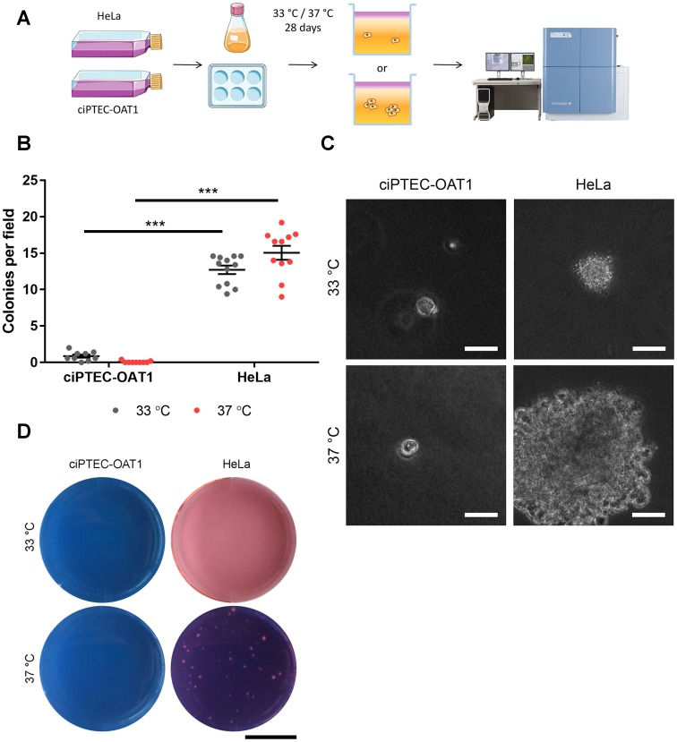
Figure 3. Anchorage-independent growth at permissive and non-permissive temperatures.
(A) Schematic diagram of soft agar assay. Single ciPTEC-OAT1 and HeLa cells were seeded in agarose-containing medium (0.3% (w/v)) and incubated for 28 days at either 33° C or 37° C. Cell growth and colony formation was detected by confocal imaging. (B) Quantification of colonies detected for ciPTEC-OAT1 and HeLa cells presented as number of colonies per field. (C) Representative microscopic pictures of cell colonies formed by ciPTEC-OAT1 HeLa cells after 28 days culture at 33° C and 37° C. Scale bars denote 50 μm. (D) Representative macroscopic pictures of ciPTEC-OAT1 and HeLa cell colonies. Scale bar denotes 1 cm. Values are expressed as the mean ± SEM of three independent experiments performed in triplicate. *** p < 0.001 (unpaired two-tailed Student’s t-test).

