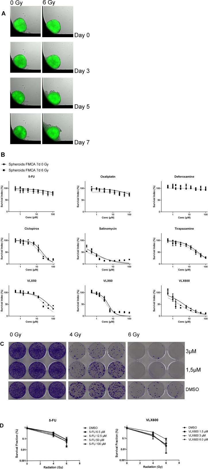
Figure 1.
(A) HCT116 GFP cells cultured as spheroids for 7 days, irradiated day 0 and then analyzed. Control spheroids (left column) vs irradiated spheroids (right column). The spheroids were typically 400–500 μm in diameter at day 0. (B) Cell survival in the FMCA assay, expressed as SI of HCT116 GFP cells cultured as spheroids for 7 days, then incubated with drugs for 7 days with irradiation (6 Gy) at 4–6 h after addition of drug. Mean ± SEM based on 3–7 independent experiments, with duplicate wells for each drug concentration. (C) Clonogenic assay with VLX600, shown as growth of HCT116 GFP cells cultured as spheroids for 7 days, then irradiated (6 Gy) 4–6 h after drug addition and 20 h later dissociated into single cells, transferred to 6-well plates and incubated for 10 days. Triplicate wells for each drug concentration. (D) Cell survival in the clonogenic assay, expressed as survival fraction of HCT116 GFP cells cultured as spheroids for 7 days, then irradiated (6 Gy) 4–6 h after drug addition and 20 h later dissociated into single cells, transferred to 6-well plates and incubated for 10 days. Mean ± SEM based on 2–3 independent experiments, with triplicate wells for each drug concentration.

