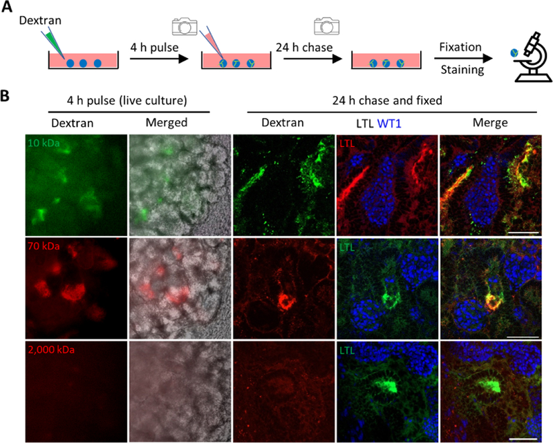
Figure 4. In Vitro Functional Validation of 3D Kidney Organoids.

(A) Schematic of in vitro dextran uptake assay.
(B) Left panels: live images of kidney organoids incubated with fluorescence-labeled dextran of various molecule weights. Right panels: whole-mount immunofluorescence analysis of kidney organoids following dextran update assay. Scale bars, 50 μm.
