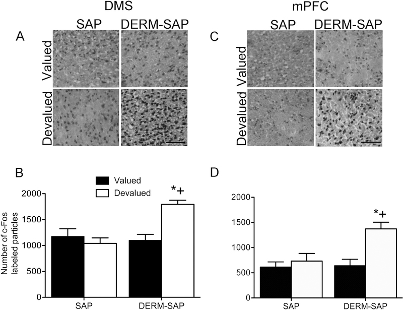
Figure 4.
Effects of patch compartment lesions in the DLS on c-Fos immunoreactivity in the DMS and mPFC (within the prelimbic subregion) following reinforcer devaluation and extinction testing. Photomicrographs showing c-Fos immunoreactivity in the DMS (A) and mPFC (C). Scale bar=100 μM. Quantitative analysis of c-Fos immunoreactivity in the DMS (B) and mPFC (D) in rats infused with SAP or DERM-SAP (17 ng/μl) in the DLS, prior to RI training, CTA and extinction testing. Data are presented as the mean (±SEM) number of c-Fos immunoreactive particles per area. *significantly different from DERM-SAP-pretreated, saline-treated rats, p<0.05; +significantly different from SAP-pretreated, LiCl-treated rats, p<0.05.

