Figure 1.

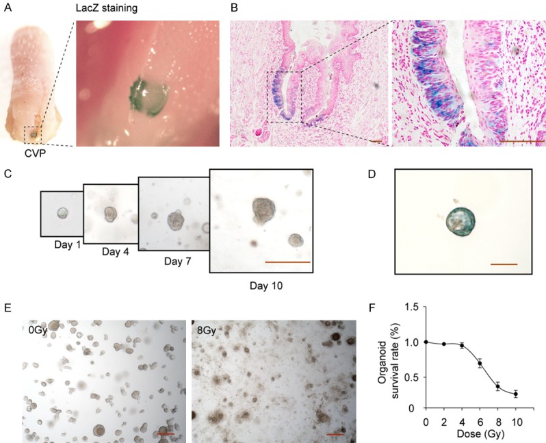
Establishment of a taste bud organoid model of radiation-induced lingual epithelial cell damage. (A) The macroscopic images of β-galactosidase-stained area (blue) in CV papilla of posterior tongue, and (B) the microscopic images of β-galactosidase stained stem cells. LacZ+ stem cells are visible at the base of the taste buds in CV papilla tissue. (C) Development of taste bud organoids in 3D cultures on days 1, 4, 7 and 10, and (D) LacZ staining. (E) radiation-induced damage of taste bud organoids, and (F) the dose curve. Dose range from 2 Gy to 10 Gy, Values are means ± SD (n=4). Scale bar 100 μm.
