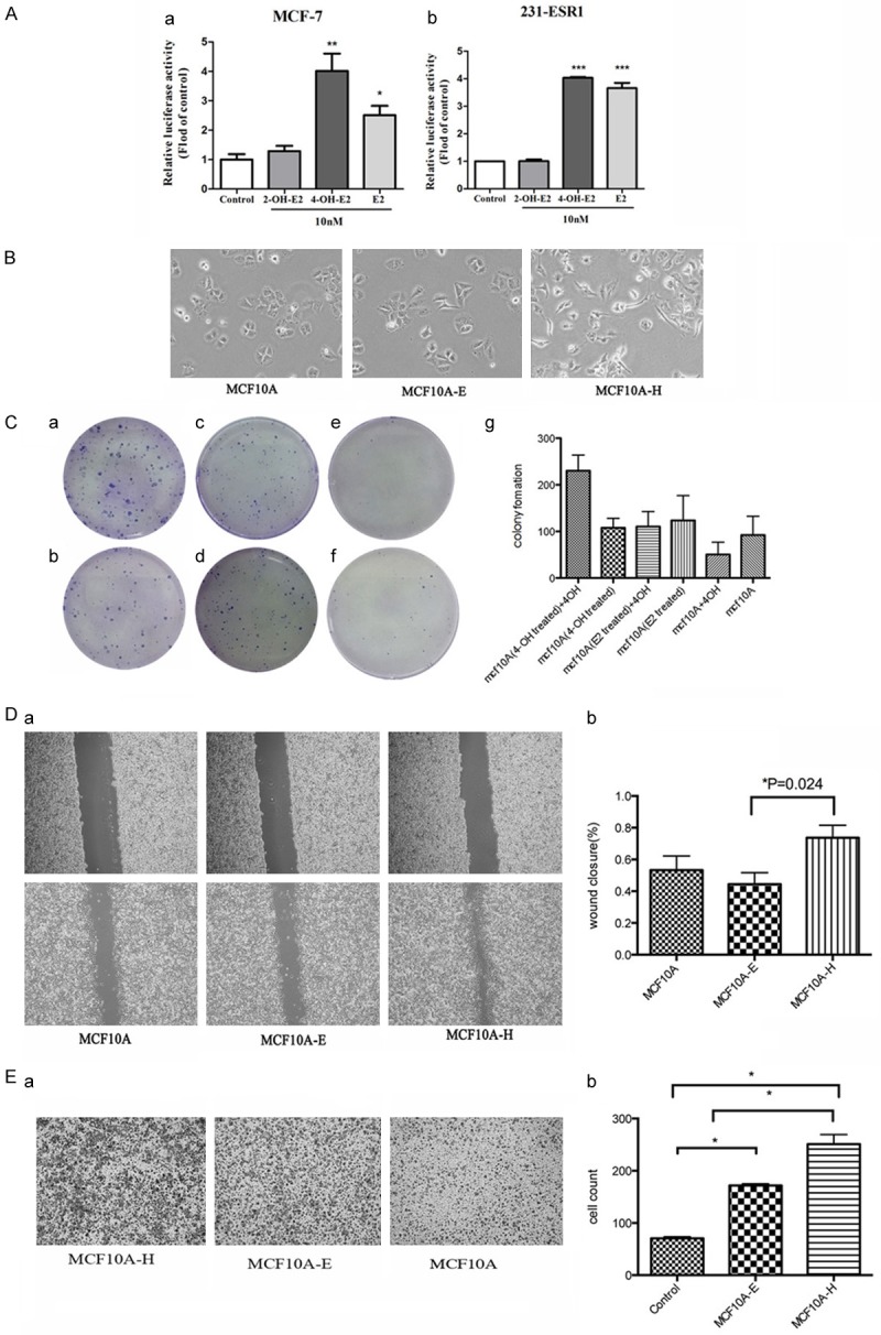
Figure 2.

(A) Estrogen-responsive luciferase reporter assay showed that 4-OH-E2 had estrogen activity. We treated MCF-7 and MDA-MB-231-ESR1 cells with 4-OH-E2, 2-OH-E2 and E2. 4-OH-E2 and E2 increased ER-α reporter activity about 4 fold over the control group. We treated MCF10A cells with 4-OH E2 (MCF10A-H) and E2 (MCF10A-E) both at 10-8 M for 2 months. (B) The effects of 4-OH E2 (MCF10A-H) and E2 (MCF10A-E) on MCF10A cells. Microscope images show that 4-OH E2 induced cell-to-cell contact and led to a higher spreading with more formation of filo podia by comparison with the E2 and control groups. (C) The cell transformation was carried out by colony formation. MCF-10A-H cells (a, b), MCF10A-E cells (c, d) and MCF10A cells (e, f) were seeded in six-well plates at a density of 500 cells/well. After 24 hours of seeding, cells were exposed to 4OH E2 (a, c, e) or not (b, d, f), The control groups were cultured in red-free DMEM media supplemented with 10% fetal bovine serum. The 4-OH E2 group was treated with 10-8 M 4-OH E2. Colony efficiency was determined by a count of the number of Colonies (more than 50 cells). The MCF10A-H cells in the culture medium with 10-8 M 4-OH E2 have much more colonies than other groups (P = 0.0013). 4-OH E2 may inhibit the growth of the MCF10A and MCF10A-E (g). (D) Cell migration was measured by using the Culture-Inserts Wound healing assay. Images of wound repair were taken at 0, 24 h after wound. The distance of wound closure is shown by area at 24 h. Representative photographs and quantification are shown, original magnification, ×200 (a), Cell migration (%) was quantified by calculating the wound width (b). (E) Transwell migration assay. Representative photographs and quantification are shown. Columns: average of three independent experiments, original magnification, ×200.
