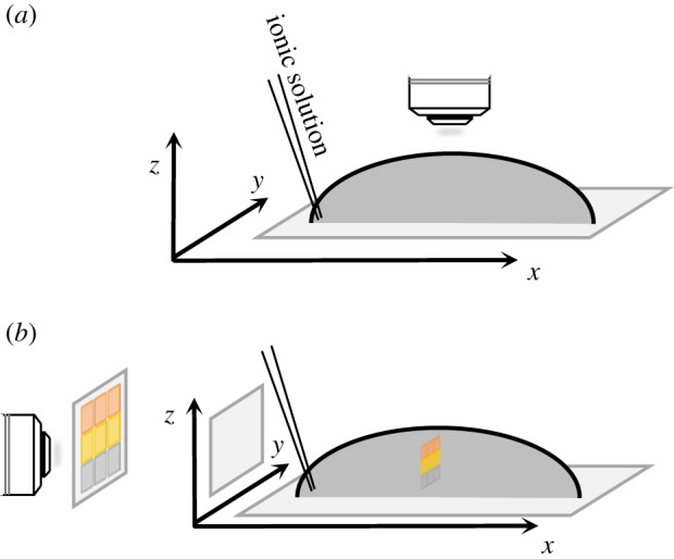
Figure 1.

Schematic views of the droplet assay. Potassium was applied at the base of each droplet containing zoospores and at a point on the circumference. Subsequent characterization of the metrics of zoospore motion was based on micrographs generated in either the horizontal (a) or vertical (b) plane. (Online version in colour.)
