Figure 4.
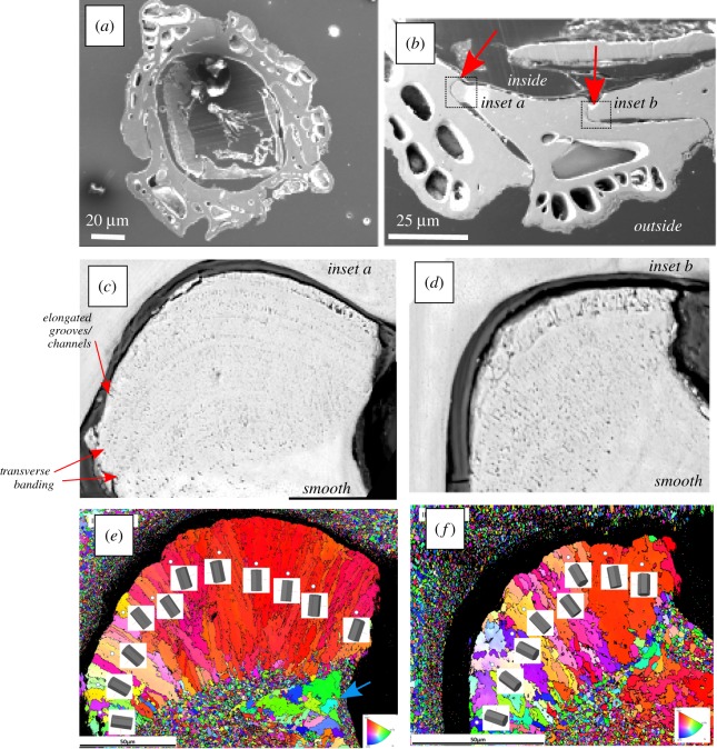
SEM imaging and EBSD crystallography of the barnacle and ala. (a) SEM image of an individual barnacle in transverse section. (b) View of three interlocking plates and ala (red arrows). (c,d) Close up of two alae (inset boxes in b), revealing microstructure transverse banding and perpendicular elongated grooves/channels at the tip. (e,f) EBSD maps of ala in (c,d), illustrating elongated grain orientations at the tip of the ala, and granular grains behind the tip and on the adjacent plate. Blue arrow illustrates inside edge coarse grains. Elongated grains appear to correlate with the porous area of the ala. Scales in (e) and (f) correspond to (c) and (d), respectively. (Online version in colour.)

