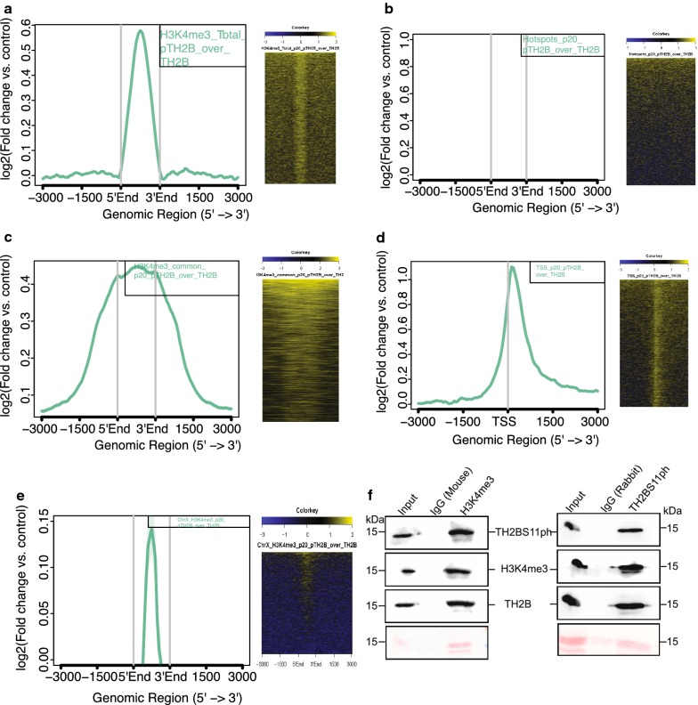
Fig. 4.
Localisation of TH2BS11ph at TSS and recombination hotspots with respect to TH2B. a Analysis of overlap of TH2BS11ph with Total H3K4me3. b Analysis of overlap of TH2BS11ph with DSB hotspots. c Overlap of TH2BS11ph with H3K4me3 common peaks representing the TSS-associated H3K4me3 marks. d Localisation of TH2BS11ph at TSS. e Overlap of TH2BS11ph with Chromosome X-associated H3K4me3 marks. The aggregation plots in a–e show the read coverage of unique TH2BS11ph reads (compared against TH2B) at ± 3 kb from the centre of the particular histone or protein marks as indicated in the boxes in the top right corner. Y-axis represents log2(fold change of TH2BS11ph versus TH2B control), X-axis represents distance in terms of kilobases. The heat map in a–e is the corresponding representation of the read coverage at ± 3 kb from the centre of occupancy of candidate histone/protein marks. f Association of H3K4me3 and TH2BS11ph in mononucleosomes. Mononucleosome IP studies to determine the coexistence of TSS-related histone mark H3K4me3 with TH2BS11ph histone modification by forward (1st panel) and reverse IP reactions (2nd panel). Ponceau stained blots have been given for reference. The first lane in all the blots represents the Input, second lane immunoprecipitation with the Mouse IgG antibody, third lane refers to the IP using the specific antibody (anti-H3K4me3 or anti-TH2BS11ph). The antibodies labelled alongside the blot refer to the antibodies that were used for western blotting

