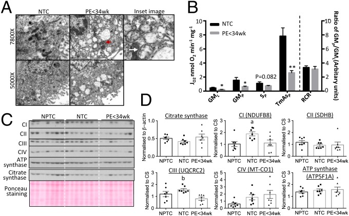
Fig. 1.
Reduction of OXPHOS capacity in mitochondria with intact ETC complexes subunits in PE < 34 wk placentas. (A) Placental mitochondria from PE appear swollen, with distorted cristae and less elongated, more rounded profiles suggestive of a high incidence of fission compared to controls. Red arrowhead indicates normal mitochondrion. (Inset ) Illustration of enlarged mitochondria with distorted cristae (arrows). The images were taken at either 5,000× or 7,800×. (B) Reduction of mitochondrial OXPHOS capacity in the PE < 34 wk placenta. Respirometry was used to measure activity of ETC complexes after addition of glutamate and malate (GML) indicating leak respiration; ADP (GMP) indicating complex I OXPHOS; rotenone + succinate (SP) indicating complex II respiration; and TMPD + ascorbate (TmAsP) corresponding to complex IV respiration. RCR was calculated as the ratio of GMP:GML. Results are presented as mean ± SEM, n for NTC = 7 and PE < 34 wk = 12. *P < 0.05; **P < 0.01. (C and D) No alteration of ETC complex subunit protein levels and constant CS in the PE < 34 wk placenta compared to NPTC. (C) Western blots. (D) Quantitative data after normalization to CS. Data are presented as mean ± SEM, n = 7. a and b indicate significant change (P < 0.05) in NPTC vs. NTC and NTC vs. PE < 34 wk, respectively. Two-tailed unpaired Student’s t test was used for statistical analysis except in D where 1-way ANOVA with Tukey’s multiple comparisons test was employed.

