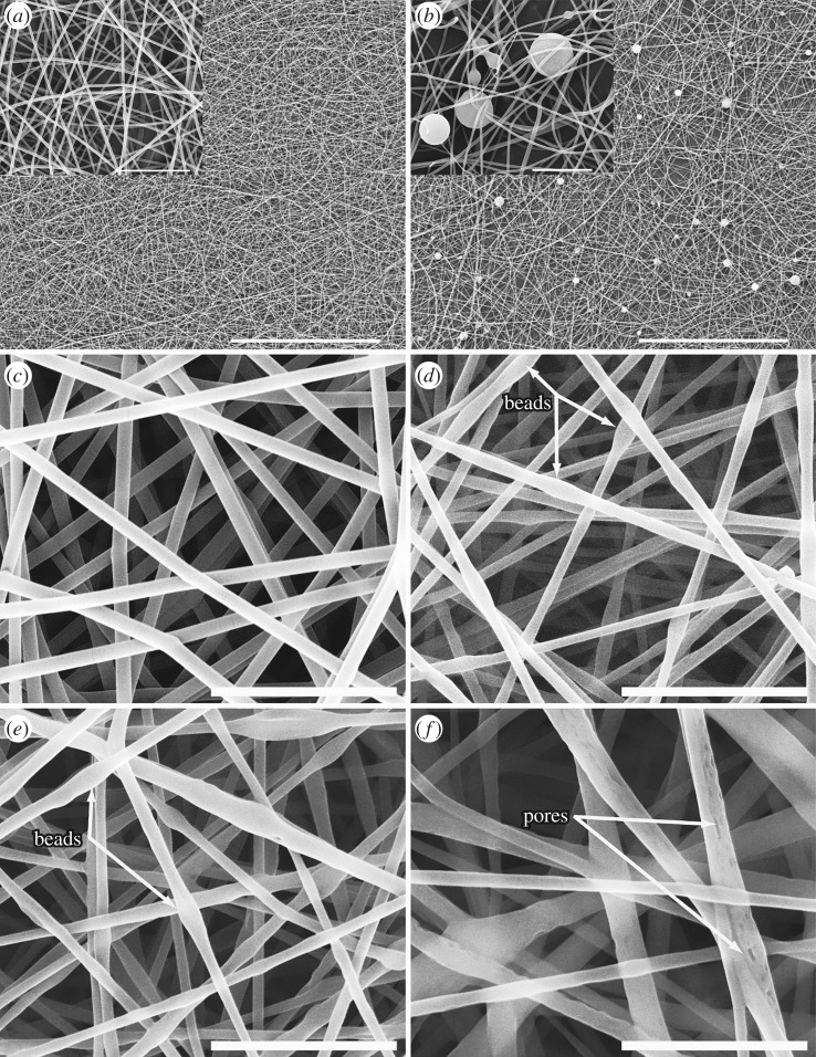
Figure 2.
SEM micrographs of PVA nanofibres: (a) blank PVA nanofibres, (b) PVA nanofibres incorporated with PMD micro-capsules, (c) blank PVA nanofibres, (d) emulsions of incorporated permethrin (8%), (e) catnip oil (8%) and (f) chilli oil (8%). Scale bar inset pictures: 100 µm for (a,b); 10 µm for (c–e); 5 µm for f. [21].

