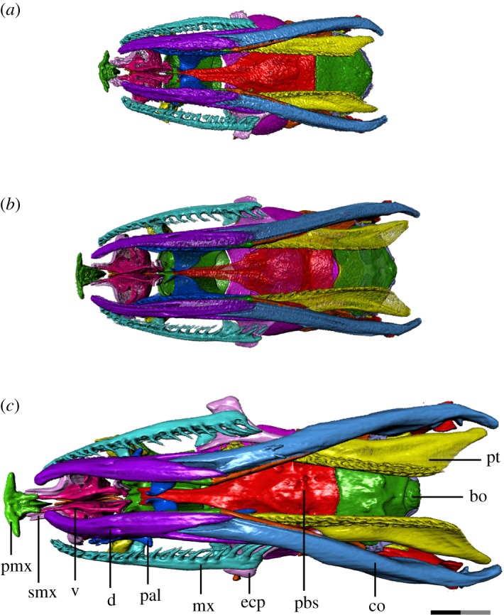
Figure 4.

Ventral view of T. radix skull throughout ontogeny: (a) embryo; (b) juvenile; (c) adult. Anterior is to the left. bo, basioccipital; co, compound bone; d, dentary; ecp, ectopterygoid; mx, maxilla; pal, palatine; pbs, parabasisphenoid; pmx, premaxilla; pt, pterygoid; smx, septomaxilla; v, vomer. Scale bar as explained in figure 1. Surface mesh (STL) files for each individually segmented bone in each stage are available in the electronic supplementary material given in [15].
