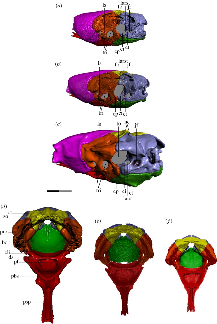
Figure 6.
Braincase of T. radix in the left posterolateral view (a–c) and anterodorsal view (d–f). Typically, colubroids (derived snakes) possess a CCF in which the cristae prootica, interfenestralis and tuberalis are elaborated into a shelf that partially covers the stapedial footplate/fenestra ovalis; however, in T. radix, this feature is conspicuously absent. (a,f) Embryo; (b,e) juvenile; (c,d) adult. In both a and b, note the lack of fusion between the cristae interfenestralis and tuberalis, leaving the LARST open laterally. bo, basioccipital; cli, clinoid process; ci, crista interfenestralis; cp, crista prootica; ct, crista tuberalis; ds, dorsum sellae; fo, fenestra ovalis; jf, jugular foramen; larst, lateral aperture of the recessus scalae tympani, ls, laterosphenoid; nc, nuchal crest; ot, otoccipital; pbs, parabasisphenoid; pf, pituitary fossa; pro, prootic; psp, parasphenoid process of the parabasisphenoid; so, supraoccipital; tri, trigeminal openings. Note: scale bar as explained in figure 1. Surface mesh (STL) files for each individually segmented bone in each stage are available in the electronic supplementary material given in [15].

