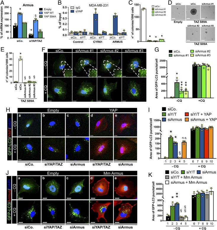
Fig. 5.
YAP/TAZ control autophagic flux through their direct target Armus. (A) MII-GFP-LC3, infected with an empty vector (empty) or with the indicated siRNA-insensitive doxycycline-inducible lentiviral YAP constructs, were transfected with control (siCo) or YAP/TAZ-targeting siRNAs (siYAP/TAZ). Cells were treated with doxycycline, harvested 48 h after siRNA transfection, and analyzed by RT-qPCR for Armus mRNA levels. Data were normalized to empty-infected cells transfected with siCo (black bar). (*P ≤ 0.0001 siCo + YAP WT compared to empty siCo; §P ≤ 0.0001 siYAP/TAZ + YAP WT compared to empty siYAP/TAZ; 2-way ANOVA). See also SI Appendix, Fig. S3 A and B. (B) Validation by ChIP-qPCR in MDA-MB-231 cells of the YAP/TAZ-binding site on the Armus-associated enhancer (see also SI Appendix, Fig. S3 C and D). CYR61 promoter is positive control, HBB promoter is negative control (control). Data from 3 replicates (mean + SD) are shown normalized to the percent input (1% of starting chromatin used as input). (C) Armus depletion impairs anchorage-independent growth. Quantification of colonies formed by MDA-MB-231 transfected with siCo or with 3 independent Armus siRNAs (siArmus #1, #2, and #3) plated in soft-agar assays. (*P < 0.0001 compared to siCo; 1-way ANOVA). See also SI Appendix, Fig. S3G for the validation of siRNAs. (D and E) Armus is required for YAP/TAZ-induced mammosphere formation. TAZ S89A-overexpressing MII cells (TAZ S89A) were transfected with the indicated siRNAs and tested for mammosphere formation. MII cells transduced with empty vector (empty) are negative control. (*P < 0.0001 TAZ S89A siCo compared to empty siCo; §P < 0.0001 TAZ S89A siArmus compared to TAZ S89A siCo; 1-way ANOVA). (F) Representative confocal images of MII-GFP-LC3 transfected as in C. The Insets (2×) show higher magnification of the GFP-LC3 puncta. (G) Quantification of GFP-LC3 puncta of MII-GFP-LC3 cells treated as in F, measured as area of GFP-LC3 puncta per cell. (*P < 0.001, **P < 0.05 compared to −CQ siCo; 2-tailed Student’s t test). (H and I) MII-GFP-LC3, infected with an empty vector (empty) or with a siRNA-insensitive doxycycline-inducible lentiviral Flag-tagged YAP constructs (YAP), were transfected with siCo, YAP/TAZ siRNAs mix (siYAP/TAZ), or Armus siRNA (siArmus), and treated with doxycycline. Representative confocal images (YAP in red) (H) and quantification (I) of GFP-LC3 puncta of MII-GFP-LC3 cells treated as above, measured as area of GFP-LC3 puncta per cell. (*P < 0.0001 compared to lane 1; §P < 0.0001 compared to lane 2; n.s., not significant compared to lane 3; 1-way ANOVA). (J) MII-GFP-LC3, infected with an empty vector (empty) or with a doxycycline-inducible vector encoding a siRNA-insensitive HA-tagged mouse Armus construct (Mm Armus, red), were transfected with the indicated siRNAs. (K) Quantification of GFP-LC3 puncta of MII-GFP-LC3 cells treated as in J. (*P < 0.01, **P < 0.001 compared to lane 1; §P < 0.001 compared to lane 2; §§P < 0.0001 compared to lane 3; 1-way ANOVA). (F–K) Cells were treated with medium (−CQ) or CQ 50 μM (+CQ) for the last 4 h and analyzed 48 h after siRNA transfection. (F, H, and J) GFP-LC3 (green); DAPI (blue) is a nuclear counterstain. (Scale bar, 20 μm.) (A, C, E, G, I, and K) Bars represent mean (n = 3) + SD.

