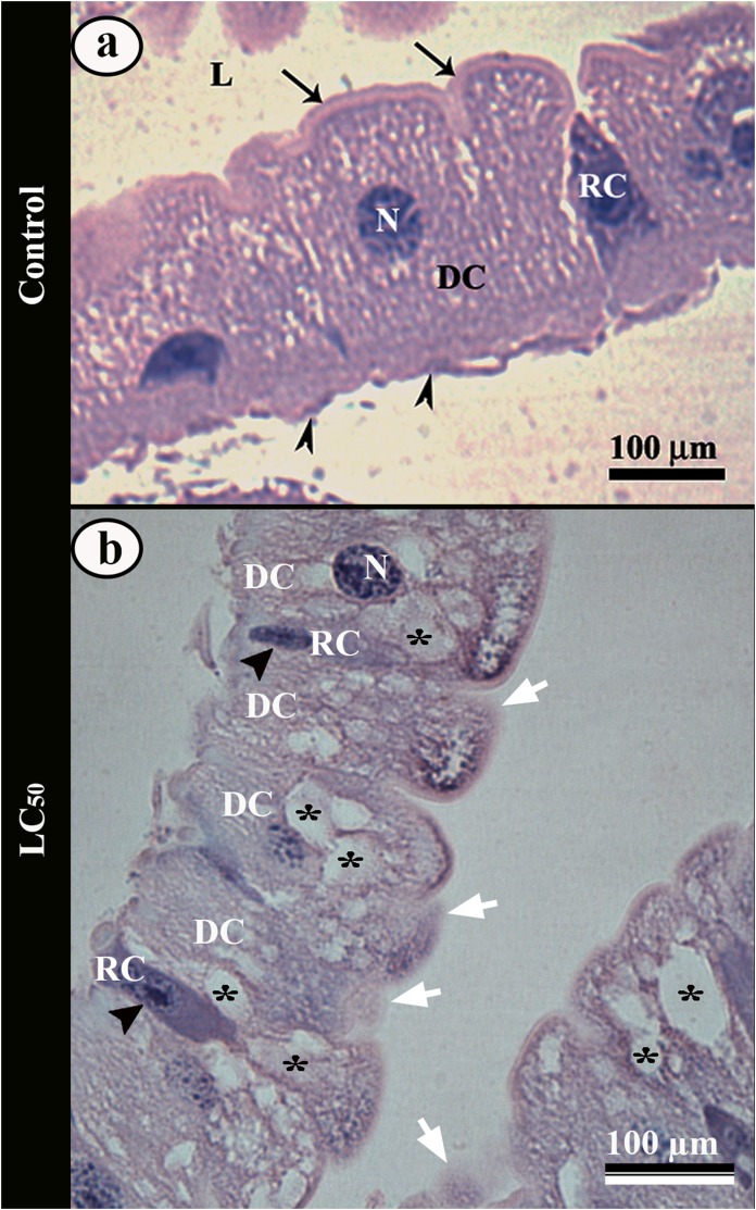
Figure 4. Light micrographs of the midgut of third instar Aedes aegypti larva.
(A) Single layered epithelium with columnar digestive cells (DC) from control larvae, with spherical nucleus (N), well-developed brush border (arrows) and basal membrane (arrowheads). Note a regenerative cell (RC) in differentiation. (B) Midgut epithelium of larvae exposed to aqueous solution of LC50 pyriproxyfen showing digestive cells (DC) with cytoplasmic vacuoles (asterisks) and disorganized brush border (arrows). Regenerative cells (RC) from the larvae exposed in concentrations. Note many regenerative cells (RC) in differentiation with large nucleus (black arrow head). L – lumen.

