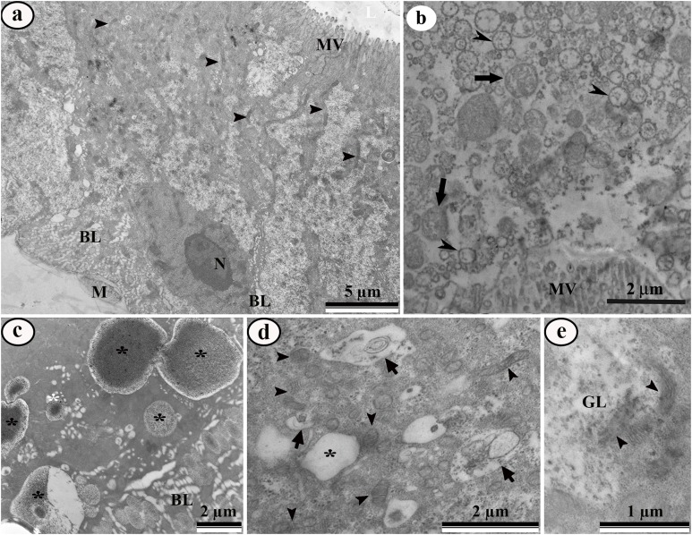
Figure 6. Transmission electron micrographs of the digestive cells from midgut of third instar Aedes aegypti larvae exposed to LC50 pyriproxyfen aqueous solution.
(A) General view showing damaged microvilli (MV), Nucleus (N), mitochondria (arrowhead) and enlarged basal labyrinth (BL) and muscle (M). (B) Midgut lumen showing cell debris similar to mitochondria (arrows) and rough endoplasmic reticulum (arrowheads). (C) Basal cell region showing big lipid droplets (asterisks) and enlarged basal labyrinth (BL). (D) Perinuclear cytoplasm with autophagic vacuoles (arrows), lipid droplet (asterisk) and damaged mitochondria (arrowheads). (E) Details of damaged mitochondria (arrowheads) and empty glycogen deposit (GL).

