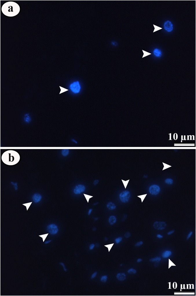
Figure 8. Micrographs of midgut epithelium of third instar Aedes aegypti larvae in the control and exposed to LC50 pyriproxyfen.
Micrographs of midgut epithelium of third instar Aedes aegypti larvae in the control (A) and exposed to LC50 pyriproxyfen (B) showing negative staining for phosphorylate histone-H3, but with increase in the number of cell nucleus (blue) in treated ones.

