Fig. 3.

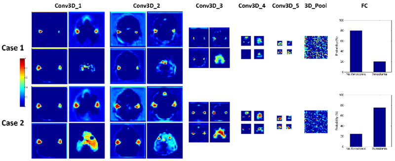
Feature map visualization. Feature maps of the first convolution layer (Conv3D_1), Conv3D_2, Conv3D_3, Conv3D_4, Conv3D_5, and 3D_Pool are presented. The 3D_Pool layer was reshaped from 1× 1 × 2 × 512 to 32 × 32 for better visualization.

Feature map visualization. Feature maps of the first convolution layer (Conv3D_1), Conv3D_2, Conv3D_3, Conv3D_4, Conv3D_5, and 3D_Pool are presented. The 3D_Pool layer was reshaped from 1× 1 × 2 × 512 to 32 × 32 for better visualization.