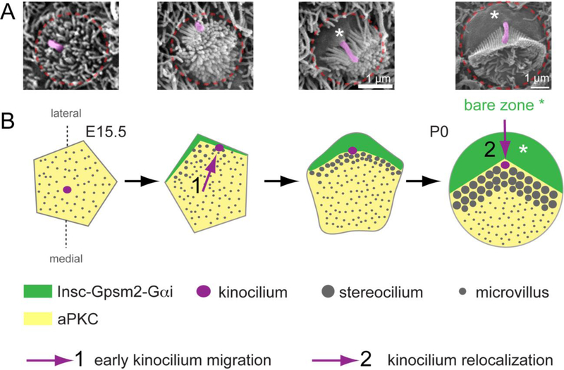
Figure 2. A molecular blueprint for planar polarization of the apical cytoskeleton.
A) SEM images of individual OHCs representative of different stages of apical differentiation. The kinocilium is highlighted in pink and the approximate OHC junction indicated in red. B) Diagram depicting changes at the HC apex from the onset of differentiation (E15.5, left) to around birth (P0, right). Initially, the aPKC kinase is uniformly enriched at the apical membrane, which is covered with microvilli, and the kinocilium occupies a central position. The first morphological evidence of planar asymmetry is the approximately lateral position of the kinocilium, which occurs at about the time the Insc-Gpsm2-Gαi complex becomes planar polarized at the lateral aspect of the cell. The Insc-Gpsm2-Gαi complex expands in surface area and labels the bare zone, the lateral region of apical membrane devoid of stereocilia or microvilli (asterisks). Insc-Gpsm2-Gαi prevents aPKC enrichment at the bare zone, establishing a molecular blueprint at the apical membrane that helps position and coordinate the hair bundle and the kinocilium. The expansion of the bare zone coincides with a relocalization of the kinocilium, from its post-migration position juxtaposed to the lateral junction to a more central position at the vertex of the chevron-shaped hair bundle around birth.

