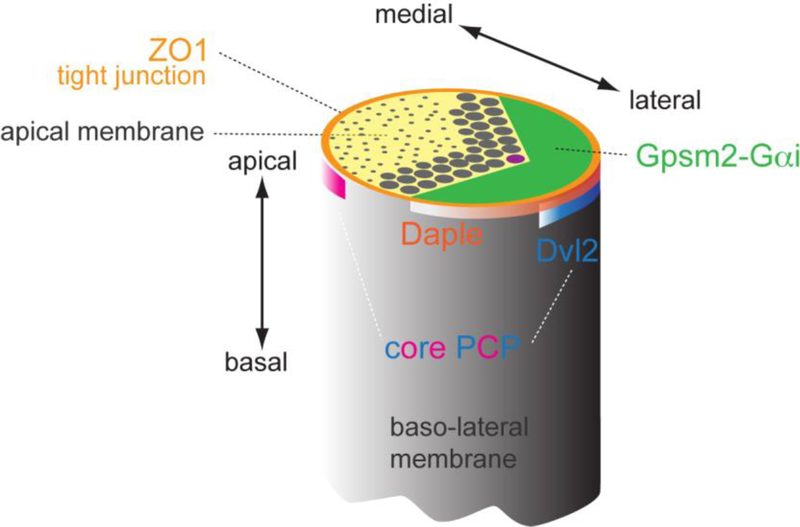
Figure 5. Distinct subcellular localizations for different polarity modules at the HC apex.
Proteins identified as planar polarized at the apical HC surface have been shown to occupy distinct subcellular domains. Core PCP proteins are enriched at adherens junctions and form antagonistic lateral (e.g. Dvl2, blue band) or medial (pink band) protein complexes. In contrast, Par3 and Daple proteins largely coincide with lateral tight junctions (orange), slightly more apical than core PCP proteins. Finally, Insc-Gpsm2-Gαi are enriched above tight junctions at the bare zone, the region of apical membrane devoid of stereocilia or microvilli (green). Close vicinity between Gpsm2-Gαi, Daple-Par3 and Dvl2 at the lateral aspect of HCs suggests that cross-talk between these protein modules and compartments may integrate intercellular PCP and cell-intrinsic cytoskeleton polarization.

