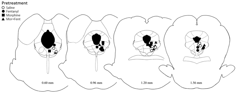
Figure 1. Location of injection sites within the vlPAG.
Cannula placements for animals pretreated with saline, morphine, fentanyl, or morphine+fentanyl. Injection sites were similar for all groups across coronal sections of the PAG. Although the image shows the location of the cannula tip, an injection volume of 0.4 μl causes the drug to diffuse into the vlPAG. Distance from Lambda are listed below each image.

