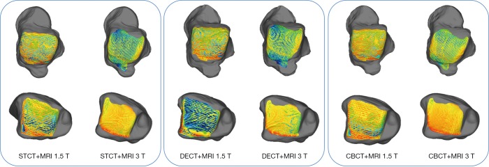
Figure 5.
Results from DMA for each combination of cartilage-to-bone models, all obtained through ICP registration: for both the talus (top row) and the tibia (bottom), the diagrams show the map of the distance between the model of the bone and the model of the individual cartilage. Bone models were from STCT (left), DECT (central), and CBCT (right); each combined with both cartilage models, i.e., from MRI 1.5 T and MRI 3 T. DMA, distance map analysis; ICP, iterative closest point; STCT, standard CT; DECT, dual energy CT; CBCT, cone-beam CT; CT, computed tomography; MRI, magnetic resonance imaging.

