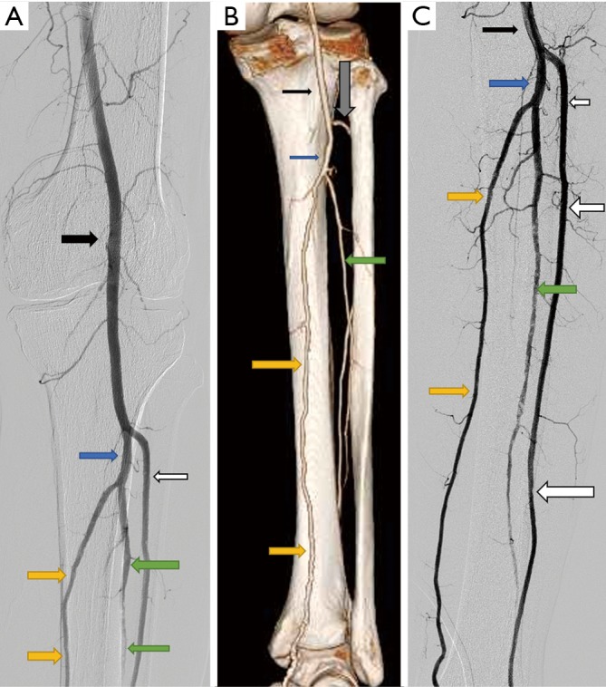
Figure 3.

Angiogram demonstrating the anatomy of popliteal and tibial arteries. (A) Digital subtraction angiogram of left leg showing a normal popliteal artery (black arrow) and its division into anterior tibial (white arrows) and tibioperoneal trunk (blue arrow). The tibioperoneal trunk further divides into peroneal artery (green arrows) and posterior tibial artery (yellow arrows); (B) CT angiogram volume rendered technique posterior view demonstrating the popliteal artery and its division. Note the site of anterior tibial artery piercing the interosseous membrane to move anteriorly (grey arrow); (C) DSA of below knee tibial vessels.
