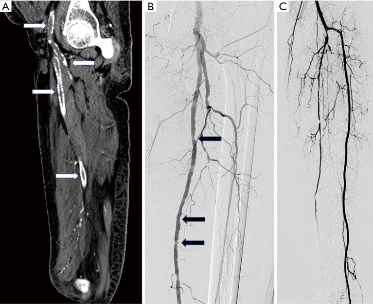
Figure 4.
Atherosclerotic disease on imaging. (A) CT plain Sagittal reformat of hip and thigh region of a 65 years old lady with poorly controlled diabetic and chronic renal failure patient showing severe vessel wall calcifications in common femoral, superficial femoral and profunda femoris artery (white arrows); (B) DSA of another 71 years old patient with diabetes and hypertension showing diffuse atherosclerotic changes in the form of multiple eccentric plaques (black arrows); (C) below knee angiogram of same patient as (B) showing single vessel runoff in the form of anterior tibial artery, peroneal artery is occluded just beyond its origin and posterior tibial artery shows diffuse disease.

