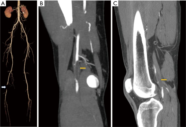
Figure 7.
Fifty-nine years old male with claudication, Cystic adventitial disease of popliteal artery (A) CT VRT showing occlusion of the right popliteal artery (white arrow), absence of atherosclerotic disease in rest of the vasculature; (B) coronal MIP of the CT angiogram showing well defined cystic lesion causing extrinsic severe compression (yellow arrow); (C) sagittal MIP of CT angiogram showing non enhancing cystic lesion causing extrinsic compression (yellow arrow).

