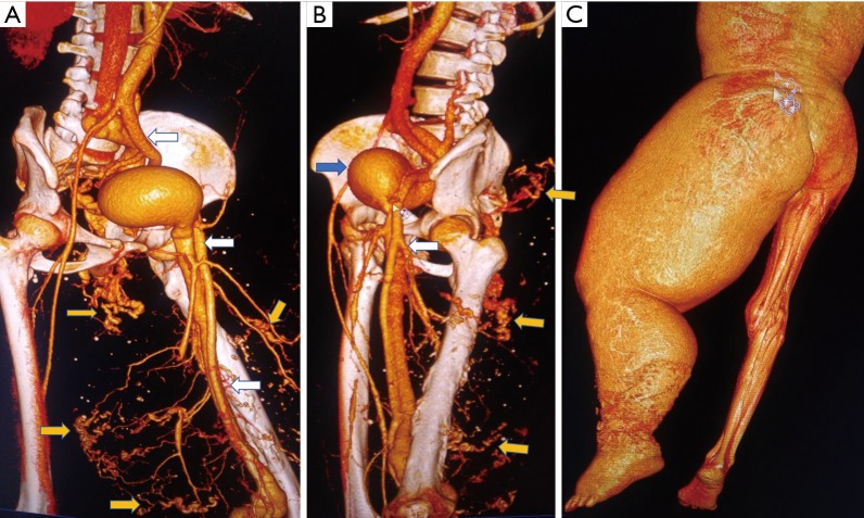
Figure 8.
CT angiogram volume rendered technique of a 31 years old male with diffuse angiomatosis of left lower limb with multiple arteriovenous malformations in the limb. (A) Dilated left lower limb arteries and veins (white arrow), note the filling of the left lower limb veins whereas the right lower limb veins are not opacified yet. The size of the left lower limb arteries is grossly enlarged compared to normal sized contralateral vessels. Multiple sites of arteriovenous malformation and arteriovenous shuntings (yellow arrow) (B) note the large venous sac in the draining vein (blue arrow); (C) grossly hypertrophied left lower limb compared to the normal right lower limb.

