Figure 9.
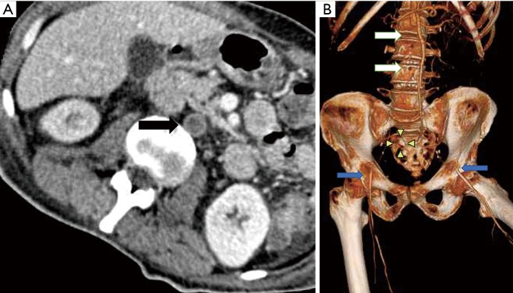
Imaging (CT) in vasculitis. (A) CT axial venous phase shows occlusion of infrarenal aorta with wall thickening and enhancement of the wall (black arrow); (B) CT angiogram VRT showing the infrarenal aortic occlusion (white arrows) with distal reformation of bilateral external iliac artery (blue arrows).

