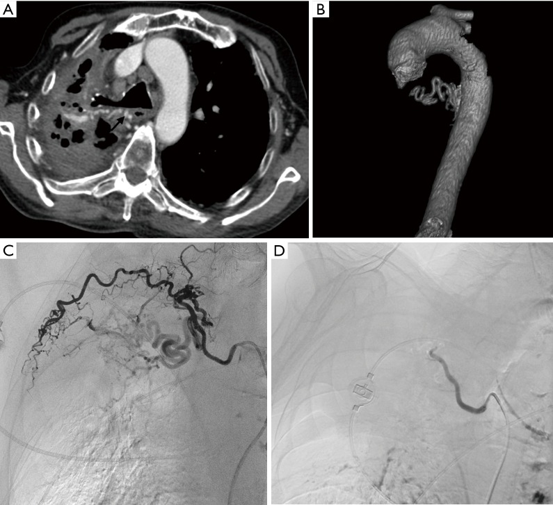
Figure 11.
A 65-year old male with massive hemoptysis and reactivated pulmonary tuberculosis. (A) extensive fibrocavitary changes of the right upper lobe noted in the axial section of the CT chest along with tortuous dilated artery (arrow) supplying the diseased lung; (B) VRT image showing the tortuous right bronchial artery; (C) DSA showed an extremely tortuous and dilated right bronchial artery supplying the diseased right upper lobe; (D) post embolisation angiogram showed stasis in the embolised bronchial artery. VRT, volume rendering technique; DSA, digital subtraction angiogram.

