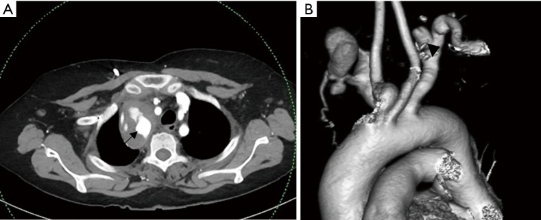
Figure 2.
Subclavian artery (SCA) aneurysm in a 56-year-old female who had history of multiple aortic and large vessel aneurysms in the past (unknown etiology). (A) Axial CT angiogram of the thorax showing the ruptured pseudoaneurysm of the right subclavian artery (arrow); (B) VRT image of the same CT apart from depicting the right SCA pseudoaneurysm, also shows residual aneurysm of the left SCA from past surgery (arrowhead). VRT, volume rendering technique.

