
Figure 11.
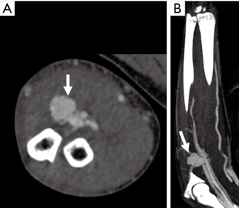
A 38-year-old man with penetrating glass injury from a motor vehicle accident 1 month ago. Axial (A) and coronal (B) CTA images showing a contrast outpouching along the proximal ulnar artery (white arrow) with extensive proximal venous enhancement along the ulnar side suggesting arteriovenous fistula. The venous enhancement starts at the pseudoaneurysm and extends proximally. CTA, CT angiography.