
Figure 17.
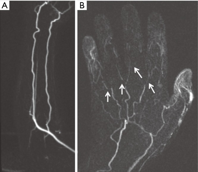
A 50-year-old woman with no previous history of any connective tissue disorder and symptoms consistent with “Raynaud phenomenon”. Subtracted coronal time-resolved MRA MIP images of the forearm (A) and hand (B) show normal forearm arteries with non-visualization of the superficial palmar arch, small caliber metacarpal arteries with multifocal narrowing, and non-visualization of the digital branches (arrows) consistent with “Raynaud Disease”. MIP, maximum intensity projection. Reproduced with permission from (5).