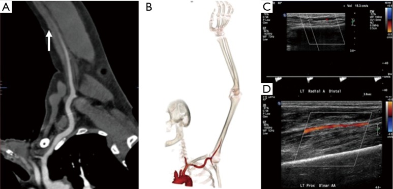
Figure 6.
A 62-year-old man with atrial fibrillation and cold left upper extremity. Curved reconstructed (A) and volume rendered (B) CT angiography images showing complete occlusion of the brachial artery in mid-arm (arrow) with non-enhancement of distal vessels. A delayed second run was not done and US Doppler images (C and D) showing slow flow in the distal vessels from collaterals or sub-total occlusion.

