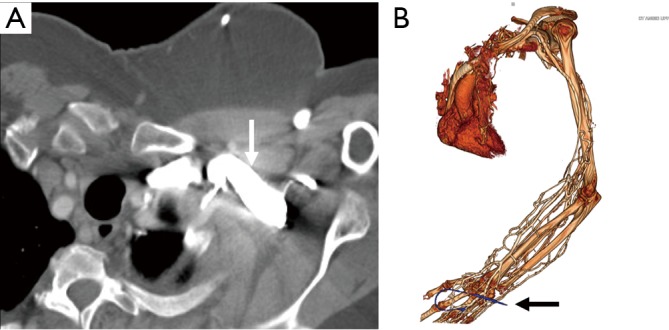
Figure 7.

CT angiography images in a 48-year-old woman with suspected vasculitis. Axial image (A) showing streak artifacts from venous contrast (white arrow) limiting arterial evaluation. Volume rendered (B) showing severe venous contamination due to contrast injection from cannula (black arrow) in the left hand.
