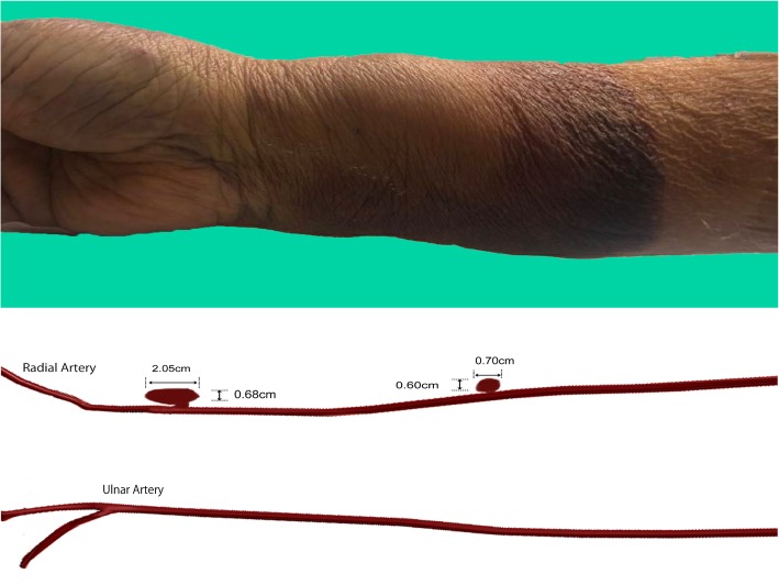
Fig. 2.
Upper panel: Day 2 after percutaneous coronary intervention, the right forearm showed swelling proximal to the puncture site. Lower panel: illustration of the location of pseudoaneurysms; the large distal pseudoaneurysm measured at 2.05 × 0.68 × 0.25 cm located near the puncture site, whereas the smaller right mid-forearm pseudoaneurysm measured at 0.70 × 0.60 × 0.09 cm located at the mid-forearm emanating from the radial artery

