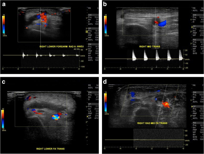
Fig. 3.
a Day 2 after percutaneous coronary intervention, duplex ultrasonography demonstrated right lower forearm pseudoaneurysm with typical “to-and-fro” waveform detected at sac neck that is consistent with a pseudoaneurysm. b Day 2 after percutaneous coronary intervention, duplex ultrasonography demonstrated right mid-forearm pseudoaneurysm with positive and negative blood flow shown on duplex Doppler. c After 2 days of TR band compression, duplex ultrasonography of the right lower forearm showed complete thrombosed pseudoaneurysm with no color Doppler signals inside. d Four days after discharge, duplex ultrasonography of the right mid-forearm showed completely thrombosed pseudoaneurysm

