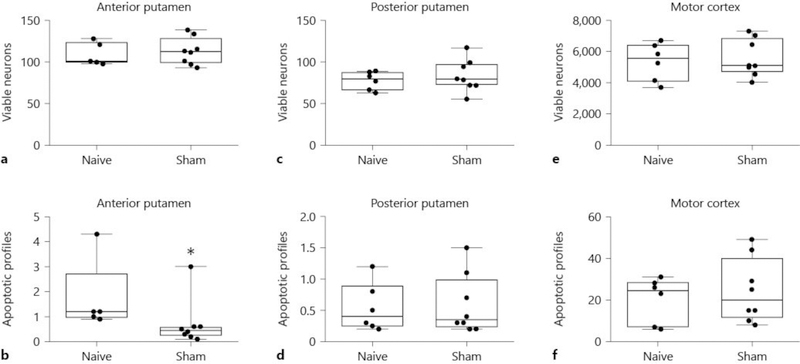
Fig. 4.
Viable neuron and apoptotic profile counts in anterior (a, b) and posterior (c, d) putamen and in motor cortex (e, f) of naïve and sham pigs. Counts were made on hematoxylin and eosin (H&E)-stained sections. The viable neuron counts were similar in naïve unanesthetized and sham normothermic piglets (a, p = 0.693 for anterior putamen; (c), p = 0.311 for posterior putamen). Naïve piglets had more apoptosis than did sham piglets, with a difference in medians of 0.75 apoptotic profiles per microscope field between groups in anterior putamen (b, * p < 0.05). Apoptotic profile counts did not differ in posterior putamen (d, p = 1.00). e, f Viable neuron and apoptotic profile counts did not differ in the motor cortex. Each circle represents 1 piglet. Box plots with IQRs and 5–95th percentile whiskers are shown.

