Figure 1.

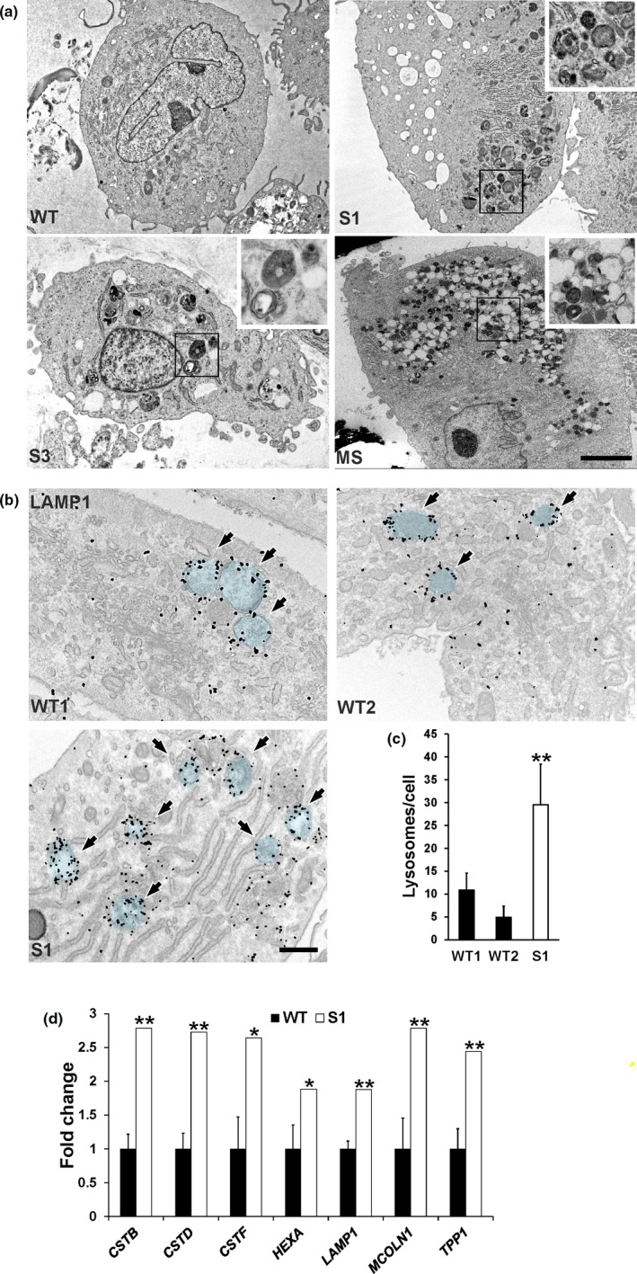
Lysosomal storage inclusions in GPHYSD fibroblasts. (a) Representative EM images of cultured skin fibroblasts from patients with GPHYSD [subject 1 (S1) and subject 3 (S3], Myhre syndrome (MS) carrying the p. (Arg496Cys) variant in SMAD4 gene showing intracytoplasmic multilamellar and electron‐dense inclusions in GPHYSD and MS fibroblasts. Cell from healthy subject (WT) is shown as control. Scale bar: 5 µm. (b) LAMP‐1 immunogold staining in subject 1 fibroblasts (S1) showing storage within LAMP‐1 decorated lysosomal vesicles highlighted in blue and indicated by arrows. Cells from two healthy subjects (WT1 and WT2) are shown as controls. Scale bar: 500 nm. (c) LAMP‐1–positive vesicle count in GPHYSD fibroblasts (subject 1, S1) compared to two WT controls (WT1 and WT2). Averages ± SEM are shown; **p < 0.005 versus WT1 and WT2; t test. (d) qPCR for lysosomal genes in GPHYSD fibroblasts from subject 1 compared to WT controls (n = 4). Averages ± SEM are shown; *p < 0.05, **p < 0.005; one sample t test
