Figure 2.

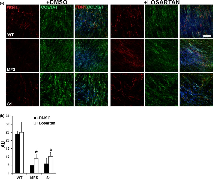
Losartan improves microfibril deposition defect in GPHYSD fibroblasts. Fibroblasts from GPHYSD and MFS patients and from healthy controls (WT) were treated for 14 days with vehicle (DMSO) or 200 μM losartan. (a) Staining for FBN1 (red) revealed improved microfibril deposition in losartan‐treated GPHYSD (subject 1, S1) and MFS fibroblasts carrying the p.Asp2104Ter variant in FBN1, compared to vehicle‐treated (DMSO) cells. COL1A1 (green) staining is shown as ECM deposition control and appears to be not affected by treatment. Nuclei were counterstained with DAPI (blue). Magnification: 20×. Scale bar: 100 μm. (b) Quantification of fluorescence intensity showing significant increase in FBN1 microfibrils deposition after losartan treatment in GPHYSD and MFS fibroblasts. Ratio between FBN1 fluorescence intensity and fluorescence area is expressed as arbitrary units (AU). Averages ± SEM are shown; *p < 0.05; t test
