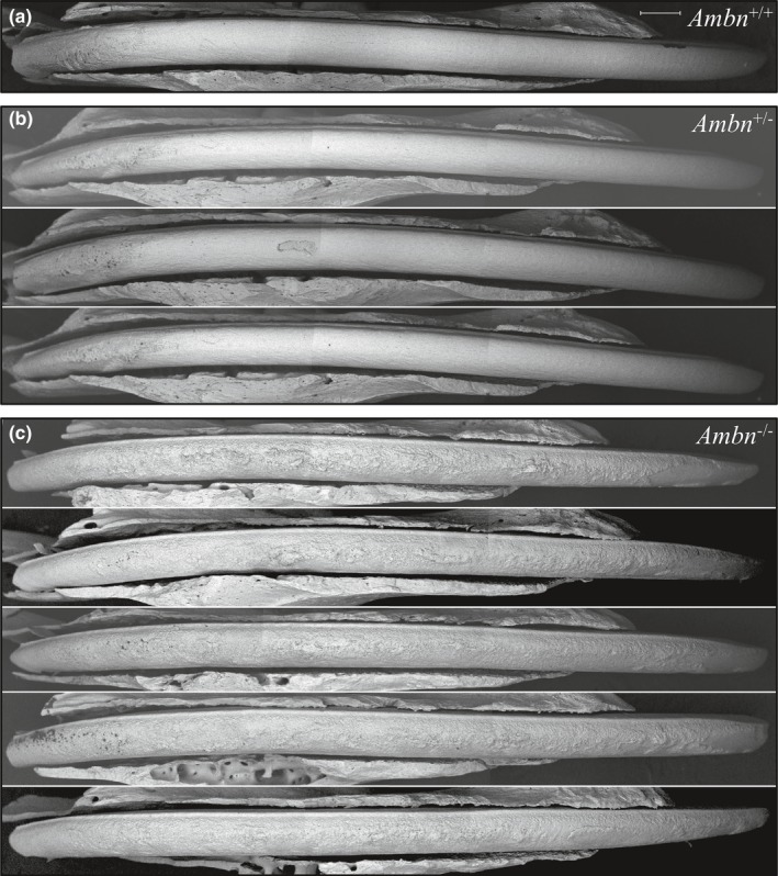
Figure 7.

bSEM montages of 7‐week mandibular incisors denuded of overlying tissues. The labial surfaces are shown, from the basal (left) to erupted (right) ends of the incisors. Dark areas contain less surface mineral than white areas. The surface of secretory stage enamel (left ~1/4 of the incisor length) appears rough in all genotypes. The maturation stage enamel surface of the Ambn +/+ incisor (a) was similar to those from the Ambn +/lacZ mice (b) whereas the Ambn lacZ/lacZ homozygous incisors (c) exhibited very rough, poorly mineralized (dark), crusty surfaces with protruding nodules. Scale bar = 500 µm
