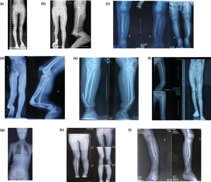
Figure 2.

The radiological evidence of NF1 in our patients. (a) II‐1 of family 1: the radiograph demonstrates her congenital pseudarthrosis of the left tibia. (b) II‐2 of family 2: the radiographs demonstrate his congenital pseudarthrosis of the right tibia. (c) III‐2 of family 3: the radiographs demonstrate her congenital pseudarthrosis of the left tibia. (d) III‐6 of family 4: the radiographs demonstrate his congenital pseudarthrosis of the right tibia. (e) III‐3 of family 5: the radiographs demonstrate her congenital pseudarthrosis of the left tibia. (f) III‐2 of family 7: the radiographs demonstrate his unequal leg length deformity. (g) II‐2 of family 8: the radiograph demonstrates his scoliosis. (h) III‐2 of family 9: the radiograph demonstrates her tibial bowing deformity. (i) IV‐1 of family 3: the radiographs demonstrate her congenital pseudarthrosis of the left tibia. All of the images are published with permission from the affected individuals
