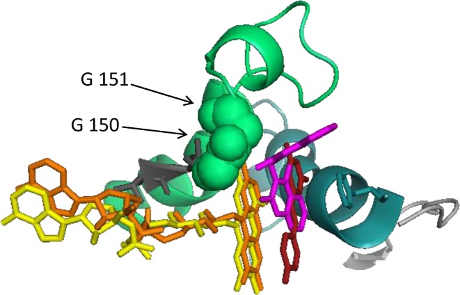
Figure 3. Structural alignment of NQO1 with NQO2.
Dicoumarol is shown in pink, resveratrol in red, FAD from NQO2 in orange and FAD from NQO1 in yellow. Resveratol is flat and does not make contact with the glycine residues at position 150 and whereas dicoumarol is bent and does make contact with these residues. The alignment was made using PyMol (www.pymol.org), the command used aligns all atoms but includes an outlier rejection to ignore parts that deviate by more than 2 Å. Structures were taken from: NQO1, PDB 2F1O [11] with FAD from PDB 1QBG [9] and resveratrol from PDB 1SG0 [61].

