Figure 2.
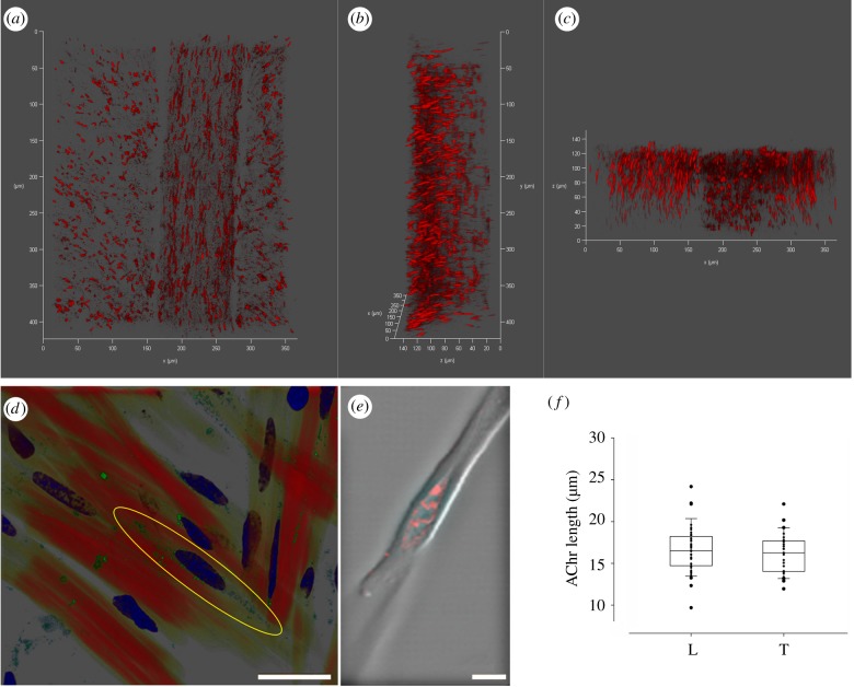
Localization of AChR in the octopus arm muscle. (a,b,c) Confocal 3D projections of an arm transverse section at 0°, 90°x and 90°y, respectively. Both L and T (trabeculae) muscles are visible and show the same longitudinal arrangement of the AChR (fluorescently labelled with α-bungarotoxin ATTO-633). (d) Confocal image of arm transverse section (T muscles) showing single muscle cells in red (F-actin labelled with phalloidin), AChR in green (fluorescently labelled with Alexa Fluor 488 α-bungarotoxin) and nuclei in blue (DAPI). The localization of the AChR in a central area around the nucleus is highlighted by a yellow ellipse. (e) Tetramethylrhodamine-α-bungarotoxin labelling of AChR in an isolated muscle cell as seen under a confocal microscope. AChR are concentrated in an eye-shaped structure at a single site forming hill-like structures in a central area around the nucleus. (f) Length of the AChR in longitudinal (L) and transverse (T) muscles show no significant differences (t-test, p = 0.42). Scale bars: (d) 25 µm; (e) 5 µm.

