Figure 3.
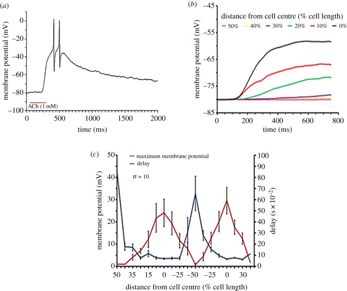
Physiological characterization of the central localization of AChR in octopus muscle cells. (a) Application of a few nanolitres of ACh (1 mM) close to the cell caused depolarization that could reach the APs threshold. (b) Typical recordings showing the membrane potential response to ACh injection at different distances along the cell. The distance with respect to the cell's centre is shown in % of total cell length. (c) Membrane potential response and the delays from ACh application as a function of the relative distance (% of cell length) of ACh injection site from the centre of the muscle cell. Each experiment was performed on a single cell. In each experiment, the ACh injecting pipette was moved from one end of the cell (indicated by 50%) to the other (indicated by −50%) and back. The results are reported as average ± s.e.m. (n = 10). (Online version in colour.)

