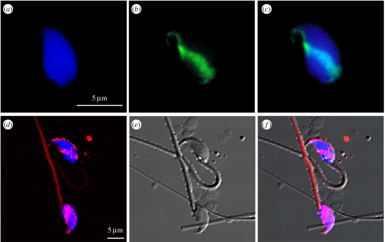
Figure 1.
Exogenous plasmid DNA and human exosomes are taken up by mouse epididymal spermatozoa: (a) DAPI staining of sperm DNA; (b) subacrosomal localization of foreign plasmid DNA revealed by FISH analysis; (c) merge of the DAPI and FISH images. Confocal microscopic images of: (d) murine spermatozoa incubated for 2 h with rhodamine-stained human exosomes (red hue) and nuclei counterstained with Hoechst (blue hue); (e) spermatozoa morphology visualized by differential interference contrast (DIC); (f) merged Rhodamine/Hoechst/DIC signals. Exosomes were extracted from A-375 human melanoma cell line as in [36]. The exosome samples shown in (d–f) were provided by courtesy of Drs S. Fais and A. Logozzi (Italian National Institute of Health). (Online version in colour.)

