Fig. 2.
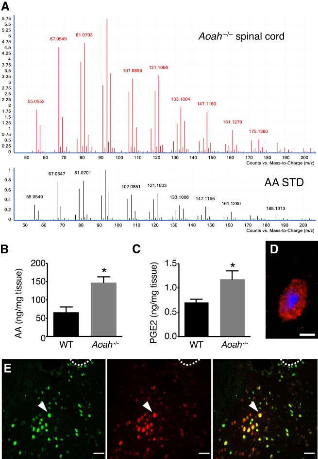
Acyloxyacyl hydrolase (AOAH)-deficient mice exhibit increased spinal arachidonic acid (AA) levels. A: MS-MS analyses of m/z 305.3 peak and AA standard reveals similar fragment spectra. B: lipidomic analyses in sacral spinal cords of female B6 and Aoah−/− mice shows increased AA (n = 4–6, *P = 0.0107, two-tailed Student’s t-test). C: the test showed increased PGE2 in Aoah−/− mice (n = 6, *P = 0.0329). Data are means ± SE. D: corticotropin-releasing factor immunoreactivity detected in the paraventricular nucleus (PVN) of female B6 mice. Red is CRF staining, and blue is DAPI. E: immunoreactivity of AOAH (green) and CRF (red) in brain sections of female B6 mice shows double-labeled cells (yellow) in the PVN. Dotted line, margin of the third ventricle as a landmark for the PVN.

