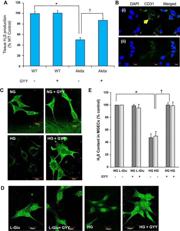
Fig. 1.
A and B: tissue H2S production and cellular H2S release in mouse glomerular endothelial cells (MGECs). A: GYY4137 (GYY) treatment increased H2S production in the kidney cortical tissue of diabetic mice. H2S was measured using 100 μl of isolated kidney cortical tissue extract from mice of different experimental groups within 36 h of sample collection. Data represent mean ± SE, n = 6 or 7/group; *, †P < 0.05. B: confocal images of MGECs fixed in 4% paraformaldehyde and labeled with or without CD31 followed by secondary goat anti-mouse IgG (Alexa Fluor 488). i: CD31-positive cells (green): yellow arrow. ii: negative control cells stained with DAPI in the absence of CD31 and presence of secondary antibody. Nuclear counterstain with DAPI showing blue color. Images were captured at ×100 magnification. C and D: GYY treatment normalized the levels of H2S in MGECs in high-glucose (HG) conditions without or with osmotic control l-Glu (25 mM). Washington State Probe-1 (WSP-1), a reactive disulfide-containing fluorescent probe, was used to detect H2S in cells. E: bar diagram represents percent of fluorescent intensity. Values are mean ± SE, n = 7/group; *, †P < 0.05. Image, original magnification ×100; scale bar, 20 µm. NG, normal glucose; WT, wild type.

