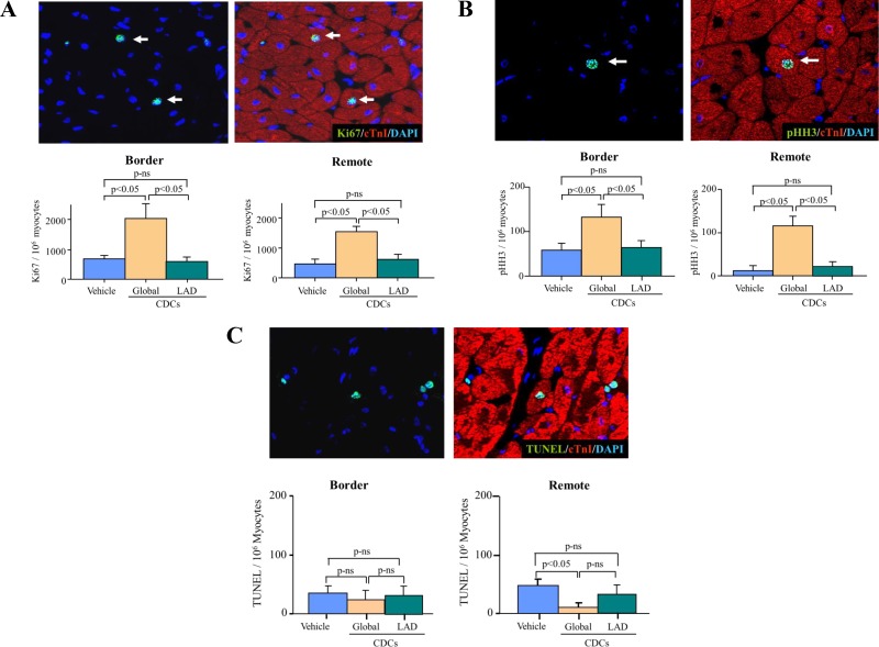
Fig. 4.
Reciprocal changes in Ki67 and phospho-Histone-H3 (pHH3) vs. myocyte apoptosis (TUNEL) in border and remote myocardium. A: four weeks after global icCDCs, Ki67-positive myocytes were significantly increased in the infarct border and remote regions of the LV vs. vehicle-treated controls with no change after LAD stop-flow icCDCs. B: cardiac myocytes in the mitotic phase (pHH3) were also increased in border and remote regions vs. vehicle-treated animals, but there was no change in remote myocardium from animals treated with LAD stop-flow icCDCs. C: global icCDCs decreased TUNEL-positive myocyte nuclei (green nuclei colocalized to myocytes with red cTnI staining) in the remote region but not in the border zone. Stop-flow icCDCs did not alter TUNEL-positive nuclei in the border or remote regions of the LV. CDC, cardiosphere-derived cells; icCDC, intracoronary cardiosphere-derived cells; LAD, left anterior descending coronary artery; LV, left ventricle; ns, not significant; TUNEL, terminal deoxynucleotidyl transferase-mediated dUTP nick end labeling.

