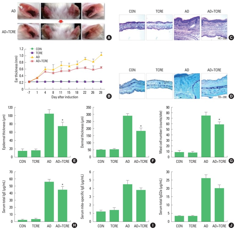
Fig. 2.
(A) Photographs of the ears of mice from each group on day 28. (B) Ear thickness was measured with a dial thickness gauge every 3 days after 2,4-dinitrochlorobenzene (DNCB) or Dermatophagoides farinae extract (DFE) application. Representative photomicrographs of ear sections stained with hematoxylin and eosin (C) or toluidine blue (D). Epidermal (E) and dermal (F) thickness was measured using the microphotographs of hematoxylin and eosin-stained tissue. (G) The number of infiltrating mast cells was determined on the basis of toluidine blue staining. Blood samples were collected by an orbital puncture on day 28. Serum total IgE (H), mite-specific IgE (I), and IgG2a (J) levels were quantified by enzyme-linked immunosorbent assay. Data are presented as the mean±standard deviation of triplicate determinations. *P<0.05, a significant difference from the value of the AD mice. AD induced by DFE and DNCB treatment. The pictures shown are representative of each group (n=3–6). The original magnification was ×100. CON, control; TCRE, tower climbing resistance exercise; AD, atopic dermatitis.

