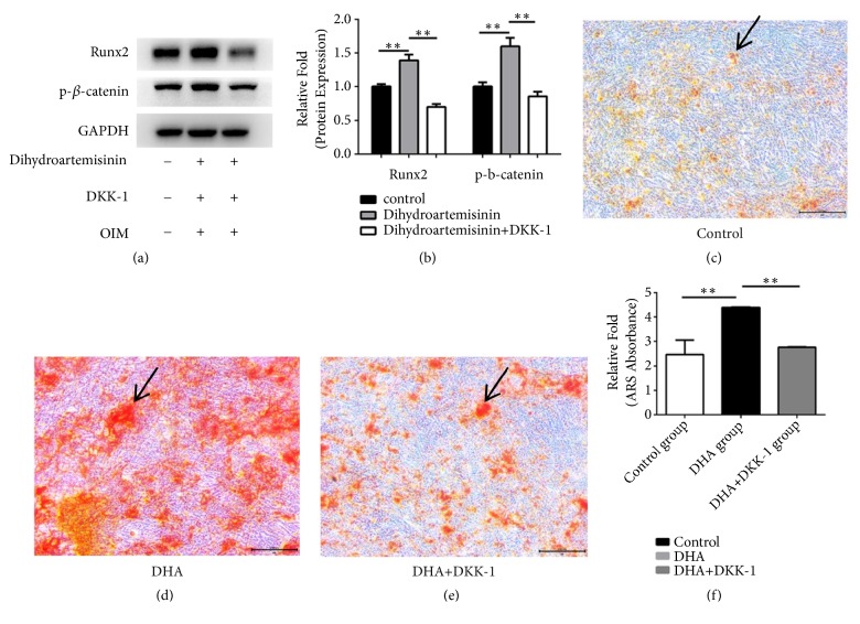
Figure 4.
(a) Human mesenchymal stem cells (hMSCs) were cultured in osteoinductive medium (OIM), OIM + 1 μM dihydroartemisinin (DHA), or OIM + 1 μM DHA + DKK-1, after which the expression levels of p-β-catenin and runt-related transcription factor 2 (RUNX2) were measured using Western blotting. (b) Protein expression levels were normalized to glyceraldehyde 3-phosphate dehydrogenase (GAPDH). ∗P < 0.05 vs. the control OIM group(c–e). hMSCs were cultured in OIM, OIM + 1 μM DHA, or OIM +1 μM DHA + DKK-1 and stained with alizarin red on day 14. (f) Mineralization was quantified by the extraction of alizarin red-stained cells. ∗P < 0.05 vs. the OIM and OIM + DHA + DKK-1 groups. Black arrows represent calcium deposits.

