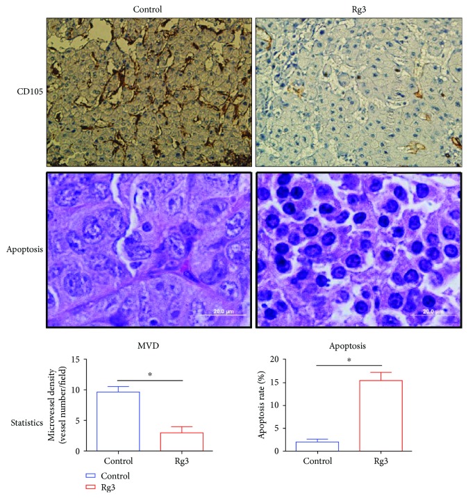
Figure 5.
The quantities of apoptotic and microvessel density analysis. Apoptotic cells and microvessels were quantified as the average of 10 fields selected per tumor (magnification was shown as the bar). The CD105 is an endothelium marker that was found highly expressed in HCC. The MVD-CD105 positively stained tumor vessels were significantly less in the Rg3 group than in the control group (P < 0.05). In addition, the apoptotic rate increased dramatically in the Rg3 group vs. the control group (P < 0.05).

